 https://smarteducation.be/wp-content/uploads/2022/08/HIITforFIT-Blog-visual-1.png
1080
1080
Robbie Billen
https://smarteducation.be/wp-content/uploads/2021/12/Logo-SmartEducation-klein-keur.png
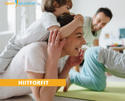
Robbie Billen2022-08-11 12:48:342024-11-12 09:01:43HIITforFIT: een efficiënt trainingsconcept bij gebrek aan tijd en energie
https://smarteducation.be/wp-content/uploads/2022/08/HIITforFIT-Blog-visual-1.png
1080
1080
Robbie Billen
https://smarteducation.be/wp-content/uploads/2021/12/Logo-SmartEducation-klein-keur.png
Robbie Billen2022-08-11 12:48:342024-11-12 09:01:43HIITforFIT: een efficiënt trainingsconcept bij gebrek aan tijd en energie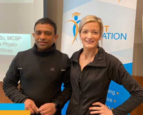 https://smarteducation.be/wp-content/uploads/2022/05/Lizzie-en-Benoy.png
1080
1080
Robbie Billen
https://smarteducation.be/wp-content/uploads/2021/12/Logo-SmartEducation-klein-keur.png
Robbie Billen2022-05-09 10:11:302024-11-12 09:02:11Lizzie Marlow & Benoy Mathew voor het eerst samen in België!
https://smarteducation.be/wp-content/uploads/2022/05/Lizzie-en-Benoy.png
1080
1080
Robbie Billen
https://smarteducation.be/wp-content/uploads/2021/12/Logo-SmartEducation-klein-keur.png
Robbie Billen2022-05-09 10:11:302024-11-12 09:02:11Lizzie Marlow & Benoy Mathew voor het eerst samen in België!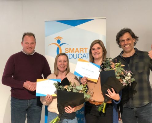 https://smarteducation.be/wp-content/uploads/2022/03/WhatsApp-Image-2022-03-11-at-2.12.15-PM.jpeg
1536
2048
Robbie Billen
https://smarteducation.be/wp-content/uploads/2021/12/Logo-SmartEducation-klein-keur.png
Robbie Billen2022-03-15 11:59:472024-11-13 13:45:57SmartEducation viert 1000e opleiding
https://smarteducation.be/wp-content/uploads/2022/03/WhatsApp-Image-2022-03-11-at-2.12.15-PM.jpeg
1536
2048
Robbie Billen
https://smarteducation.be/wp-content/uploads/2021/12/Logo-SmartEducation-klein-keur.png
Robbie Billen2022-03-15 11:59:472024-11-13 13:45:57SmartEducation viert 1000e opleiding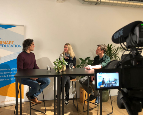 https://smarteducation.be/wp-content/uploads/2022/02/Podcast-Hoofdpijn-Aangepakt.png
1080
1080
Robbie Billen
https://smarteducation.be/wp-content/uploads/2021/12/Logo-SmartEducation-klein-keur.png
Robbie Billen2022-02-16 11:24:432024-11-12 09:06:07Hoofdpijn aangepakt: Heleen Boven en Andreas Amons aan het woord!
https://smarteducation.be/wp-content/uploads/2022/02/Podcast-Hoofdpijn-Aangepakt.png
1080
1080
Robbie Billen
https://smarteducation.be/wp-content/uploads/2021/12/Logo-SmartEducation-klein-keur.png
Robbie Billen2022-02-16 11:24:432024-11-12 09:06:07Hoofdpijn aangepakt: Heleen Boven en Andreas Amons aan het woord! https://smarteducation.be/wp-content/uploads/2022/01/Thumbnail-Catherine-Goedgezelschap-1080-x-1080-px-1-2.png
1080
1080
Robbie Billen
https://smarteducation.be/wp-content/uploads/2021/12/Logo-SmartEducation-klein-keur.png
Robbie Billen2022-01-31 10:22:412024-11-12 09:06:39Topsport combineren met kinesitherapie? Michael Somers getuigt!
https://smarteducation.be/wp-content/uploads/2022/01/Thumbnail-Catherine-Goedgezelschap-1080-x-1080-px-1-2.png
1080
1080
Robbie Billen
https://smarteducation.be/wp-content/uploads/2021/12/Logo-SmartEducation-klein-keur.png
Robbie Billen2022-01-31 10:22:412024-11-12 09:06:39Topsport combineren met kinesitherapie? Michael Somers getuigt! https://smarteducation.be/wp-content/uploads/2022/01/studentenaanbod-cover.jpg
500
750
Robbie Billen
https://smarteducation.be/wp-content/uploads/2021/12/Logo-SmartEducation-klein-keur.png
Robbie Billen2022-01-26 14:46:502024-11-12 09:07:05Postacademische vorming voor studenten
https://smarteducation.be/wp-content/uploads/2022/01/studentenaanbod-cover.jpg
500
750
Robbie Billen
https://smarteducation.be/wp-content/uploads/2021/12/Logo-SmartEducation-klein-keur.png
Robbie Billen2022-01-26 14:46:502024-11-12 09:07:05Postacademische vorming voor studenten https://smarteducation.be/wp-content/uploads/2022/01/pelvic-rehab-women-2.png
1080
1080
Robbie Billen
https://smarteducation.be/wp-content/uploads/2021/12/Logo-SmartEducation-klein-keur.png
Robbie Billen2022-01-25 08:55:252024-11-12 09:07:29Samen kunnen we de problematiek normaliseren en het taboe doorbreken!
https://smarteducation.be/wp-content/uploads/2022/01/pelvic-rehab-women-2.png
1080
1080
Robbie Billen
https://smarteducation.be/wp-content/uploads/2021/12/Logo-SmartEducation-klein-keur.png
Robbie Billen2022-01-25 08:55:252024-11-12 09:07:29Samen kunnen we de problematiek normaliseren en het taboe doorbreken! https://smarteducation.be/wp-content/uploads/2022/01/praktijk-vehuren-vennootschap-e1641475772460.jpeg
900
900
Robbie Billen
https://smarteducation.be/wp-content/uploads/2021/12/Logo-SmartEducation-klein-keur.png
Robbie Billen2022-01-10 12:20:172024-11-12 09:07:56Je praktijk verhuren aan je vennootschap: een goed idee?
https://smarteducation.be/wp-content/uploads/2022/01/praktijk-vehuren-vennootschap-e1641475772460.jpeg
900
900
Robbie Billen
https://smarteducation.be/wp-content/uploads/2021/12/Logo-SmartEducation-klein-keur.png
Robbie Billen2022-01-10 12:20:172024-11-12 09:07:56Je praktijk verhuren aan je vennootschap: een goed idee?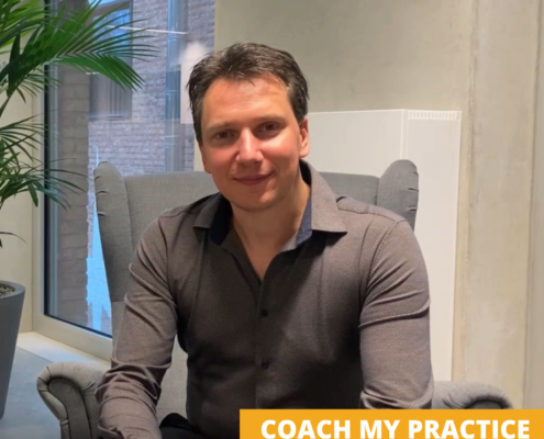 https://smarteducation.be/wp-content/uploads/2022/01/Thumbnail-Catherine-Goedgezelschap-1080-x-1080-px-1.png
1080
1080
Robbie Billen
https://smarteducation.be/wp-content/uploads/2021/12/Logo-SmartEducation-klein-keur.png
Robbie Billen2022-01-07 11:44:552024-11-12 09:08:39Coach My Practice: jouw vertrekpunt én versnelling als zorgondernemer!
https://smarteducation.be/wp-content/uploads/2022/01/Thumbnail-Catherine-Goedgezelschap-1080-x-1080-px-1.png
1080
1080
Robbie Billen
https://smarteducation.be/wp-content/uploads/2021/12/Logo-SmartEducation-klein-keur.png
Robbie Billen2022-01-07 11:44:552024-11-12 09:08:39Coach My Practice: jouw vertrekpunt én versnelling als zorgondernemer!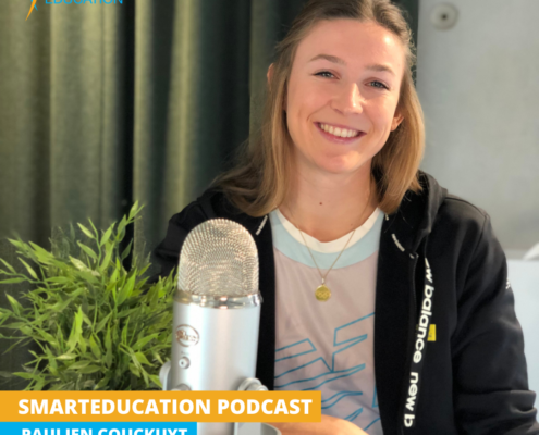 https://smarteducation.be/wp-content/uploads/2021/12/SMARTEDUCATION-PODCAST.png
1080
1080
Robbie Billen
https://smarteducation.be/wp-content/uploads/2021/12/Logo-SmartEducation-klein-keur.png
Robbie Billen2021-12-21 09:59:062021-12-21 09:59:06Topsport en kinesitherapie: Paulien Couckuyt maakt de combinatie!
https://smarteducation.be/wp-content/uploads/2021/12/SMARTEDUCATION-PODCAST.png
1080
1080
Robbie Billen
https://smarteducation.be/wp-content/uploads/2021/12/Logo-SmartEducation-klein-keur.png
Robbie Billen2021-12-21 09:59:062021-12-21 09:59:06Topsport en kinesitherapie: Paulien Couckuyt maakt de combinatie!